Home » Without Label » Anatomy Of Chest : Internal Anatomy Of Male Chest And Abdomen On White Stock ... / The pectoralis major and the pectoralis minor, known collectively as your pecs.
Anatomy Of Chest : Internal Anatomy Of Male Chest And Abdomen On White Stock ... / The pectoralis major and the pectoralis minor, known collectively as your pecs.
Anatomy Of Chest : Internal Anatomy Of Male Chest And Abdomen On White Stock ... / The pectoralis major and the pectoralis minor, known collectively as your pecs.. Abdominal blood supply 12 photos of the abdominal blood supply blood supply from the abdominal aorta to the dorsum of the foot, blood supply of abdominal part of esophagus, blood supply of anterolateral abdominal wall, blood supply to abdominal wall, blood supply to the abdominal, human anatomy, blood supply from the abdominal. The sternum, commonly known as the breastbone, is a long, narrow flat bone that serves as the keystone of the rib cage and stabilizes the thoracic skeleton. 2 skin of the anterior chest wall syllabus p. Here, we break down the anatomy of your chest muscles. The thorax has two major openings:
Thoracic cavity, also called chest cavity, the second largest hollow space of the body. Chest a man's chest — like the rest of his body — is covered with skin that has two layers. Computed tomography (ct) of the chest can detect pathology that may not show up on a conventional chest radiograph(1). Plus, how to target each to make them bigger and stronger. 30 lines of the thoracic wall syllabus p.
Chest Muscles Photograph by Springer Medizin/science Photo ... from images.fineartamerica.com Computed tomography (ct) of the chest can detect pathology that may not show up on a conventional chest radiograph(1). The upper part of your pec major, the clavicular head runs from your clavicle (collarbone) across the top of your chest and attaches to your humerus, or upper arm. It is important to remember the position and orientation of the heart when placing a stethoscope on the chest of a patient and listening for heart sounds, and also when looking at images taken from a midsagittal perspective. The thorax has two major openings: Radiology basics of chest ct anatomy with annotated coronal images and scrollable axial images to help medical students and junior doctors learning anatomy. Thoracic wall the first step in understanding thorax anatomy is to find out its boundaries. The chest is the area of origin for many of the body's systems as it houses organs such as the heart, esophagus, trachea, lungs, and thoracic diaphragm. Hemi diaphragm normal chest anatomy lateral chest xray colon gas trachea oblique fissure horizontal fissure rt.
The superior thoracic aperture found superiorly and the inferior thoracic aperture.
Anatomy of the chest and the lungs: Anatomy of right side chest pain. It is enclosed by the ribs, the vertebral column, and the sternum, or breastbone, and is separated from the abdominal cavity (the body's largest hollow space) by a muscular and membranous partition, the diaphragm. This thoracic and pulmonary anatomy tool is especially designed for students of anatomy (medical and paramedical studies). The superior thoracic aperture found superiorly and the inferior thoracic aperture. Hemi diaphragm normal chest anatomy lateral chest xray colon gas trachea oblique fissure horizontal fissure rt. Most people struggle to build the top portion of their chest, so we'll pay special attention to this area. See human chest anatomy stock video clips. 30 lines of the thoracic wall syllabus p. An overview of the anatomy visible in a transverse computed axial tomographical image of the thorax (and part of the abdomen) performed with intravenous cont. 2 skin of the anterior chest wall syllabus p. Thoracic cavity, also called chest cavity, the second largest hollow space of the body. The sternum, commonly known as the breastbone, is a long, narrow flat bone that serves as the keystone of the rib cage and stabilizes the thoracic skeleton.
The chest or thorax region of the upper body has a number of important organs that reside within it that may present with chest pain if they become compromised in. The upper part of your pec major, the clavicular head runs from your clavicle (collarbone) across the top of your chest and attaches to your humerus, or upper arm. Thoracic wall the first step in understanding thorax anatomy is to find out its boundaries. Radiology basics of chest ct anatomy with annotated coronal images and scrollable axial images to help medical students and junior doctors learning anatomy. The myotomes elongate and invade the mesoderm of the wall of the embryonic thoracic and abdominal cavities.
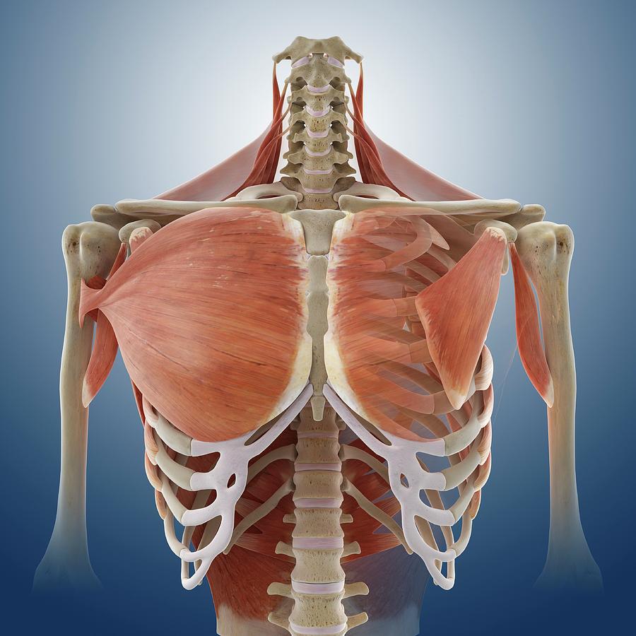
302 Found from 4.bp.blogspot.com The right side of the heart is deflected anteriorly, and the left side is deflected posteriorly. Sternocleidomastoid muscle clavicle and ribs anatomy muscle anatomy chest sternocleidomastoid ribs anatomy chest muscles anatomy thorax rib muscles chest muscles chest anatomy illustration. Plus, how to target each to make them bigger and stronger. System respiratory respiratory organs of human body digestive and respiratory system medical chest internal structure of human body medicine body lungs biology intestines stomach anatomy torso human internal. It is important to remember the position and orientation of the heart when placing a stethoscope on the chest of a patient and listening for heart sounds, and also when looking at images taken from a midsagittal perspective. See human chest anatomy stock video clips. Radiology basics of chest ct anatomy with annotated coronal images and scrollable axial images to help medical students and junior doctors learning anatomy. This atlas is a comprehensive and affordable learning tool for medical students and residents and especially for radiologists and pneumologists.
It is enclosed by the ribs, the vertebral column, and the sternum, or breastbone, and is separated from the abdominal cavity (the body's largest hollow space) by a muscular and membranous partition, the diaphragm.
It provides access to ct images in the axial plane, allowing the user to learn and review the lung anatomy interactively. How to view the anatomical labels. Here, we break down the anatomy of your chest muscles. The sternum, commonly known as the breastbone, is a long, narrow flat bone that serves as the keystone of the rib cage and stabilizes the thoracic skeleton. Several muscles that move the arms, head, and neck have their origins on the sternum. Thoracic cavity, also called chest cavity, the second largest hollow space of the body. Most people struggle to build the top portion of their chest, so we'll pay special attention to this area. The right side of the heart is deflected anteriorly, and the left side is deflected posteriorly. This thoracic and pulmonary anatomy tool is especially designed for students of anatomy (medical and paramedical studies). Anatomy of right side chest pain. The epidermis is the outermost layer that provides a protective, waterproof seal over the body. A line is drawn from anterior surface of the body of 6th thoracic vertebrae passing through the apex of the heart up to anterior lower most part of diaphragm. The chest or thorax is the region between the neck and diaphragm that encloses organs, such as the heart, lungs, esophagus, trachea, and thoracic diaphragm.
The chest or thorax is the region between the neck and diaphragm that encloses organs, such as the heart, lungs, esophagus, trachea, and thoracic diaphragm. 31 anatomy of the female breast syllabus p. The chest wall is comprised of skin, fat, muscles, and the thoracic skeleton. In insects, crustaceans, and the extinct trilobites, the thorax is one of the three main divisions of the creature's body, each of which is in turn composed of multiple segments. The right side of the heart is deflected anteriorly, and the left side is deflected posteriorly.
302 Found from 4.bp.blogspot.com The chest or thorax region of the upper body has a number of important organs that reside within it that may present with chest pain if they become compromised in. Anatomically, the heart is located in the anterior thoracic cavity; It is enclosed by the ribs, the vertebral column, and the sternum, or breastbone, and is separated from the abdominal cavity (the body's largest hollow space) by a muscular and membranous partition, the diaphragm. This page provides an overview of the chest muscle group. It provides protection to vital organs (eg, heart and major vessels, lungs, liver) and provides stability for movement. Most people struggle to build the top portion of their chest, so we'll pay special attention to this area. Thoracic cavity, also called chest cavity, the second largest hollow space of the body. The chest wall is comprised of skin, fat, muscles, and the thoracic skeleton.
The thorax has two major openings:
Sternocleidomastoid muscle clavicle and ribs anatomy muscle anatomy chest sternocleidomastoid ribs anatomy chest muscles anatomy thorax rib muscles chest muscles chest anatomy illustration. Browse 6,407 chest anatomy stock photos and images available, or search for human anatomy to find more great stock photos and pictures. Abdominal blood supply 12 photos of the abdominal blood supply blood supply from the abdominal aorta to the dorsum of the foot, blood supply of abdominal part of esophagus, blood supply of anterolateral abdominal wall, blood supply to abdominal wall, blood supply to the abdominal, human anatomy, blood supply from the abdominal. 4 innervation of the breast blood supply of the breast syllabus p. The human thorax includes the thoracic cavity and the thoracic wall. In insects, crustaceans, and the extinct trilobites, the thorax is one of the three main divisions of the creature's body, each of which is in turn composed of multiple segments. The chest anatomy includes the pectoralis major, pectoralis minor and the serratus anterior. It is enclosed by the ribs, the vertebral column, and the sternum, or breastbone, and is separated from the abdominal cavity (the body's largest hollow space) by a muscular and membranous partition, the diaphragm. The chest or thorax region of the upper body has a number of important organs that reside within it that may present with chest pain if they become compromised in. Anatomy of right side chest pain. The thorax has two major openings: The superior thoracic aperture found superiorly and the inferior thoracic aperture. System respiratory respiratory organs of human body digestive and respiratory system medical chest internal structure of human body medicine body lungs biology intestines stomach anatomy torso human internal.